 | Info
Sheets |
| | | | | | | | | | | | | | | | | | | | | | | | |
 | Out-
side |
| | | | |
|
| | | | | |  | Searchterm 'MRI scan' was also found in the following services: | | | | |
|  |  |
| |
|
(RIS) Radiology information system means a computer system that stores and processes the information for a radiology department and can be linked to the hospital information system.
The principal purpose of a RIS consists of taking over the general functions of the administration inclusive planning, monitoring and communication of all data regarding patients and its investigations in the radiology. The correct images should reach, at the correct time, the correct users. For this reason the RIS must contain a workflow management in order to simplify and steer the data flow at the individual view stations or devices (laser cameras etc.). The Radiology Information System is optimally complemented with a Picture Archiving and Communication System (PACS).
•
Collection, storage and administration of patient master data
•
Archives administration
Treatment of requirements
•
Communication (with the hospital information system, MRI scanner, other devices etc.)
| | | | | |  Further Reading: Further Reading: | Basics:
|
|
| |
| | | Searchterm 'MRI scan' was also found in the following services: | | | | |
| | |
| |
|

The range of diagnostics and imaging systems of Siemens Medical Systems covers ultrasound, nuclear medicine, angiography, magnetic resonance, computer tomography and patient monitoring. Siemens is one of the three leading MRI manufacturers, which together account for approximately 80 percent of the MRI machines installed worldwide. Siemens currently offers the Allegra 3T MRI, which is for head scanning only, but the company will also be launching the Trio MRI, a 3T whole body scanner.
Siemens has formed partnerships with more than ten research institutions and private practitioners to define a comprehensive MRI examination and compare MR to currently established cardiovascular modalities, thereby defining optimal diagnosis and treatment.
MRI Scanners:
0.2T to 1.0T:
1.5T:
3.0T to 7.0T:
Hybrid Scanners:
Mobile Solutions:
•
MAGNETOM Espree 1.5T, MAGNETOM Avanto 1.5T and MAGNETOM ESSENZA 1.5T are also offered by Siemens on certified trailers.
Contact Information MAIL
Siemens Medical Solutions
Health Services Corporation
51 Valley Stream Parkway
Malvern, PA 19355
USA | | | |
• View the DATABASE results for 'Siemens Medical Systems' (14).
| | |
• View the NEWS results for 'Siemens Medical Systems' (3).
| | | | | | Further Reading: | | Basics:
|
|
News & More:
| |
| |
| | | | | |
| |
|
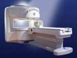
From GE Healthcare;
the Signa Ovation™ is a patient-friendly open MRI scanner designed not only to handle a typical patient mix, but to accommodate larger patients, patients who are claustrophobic, and others who have difficulty tolerating the close quarters of conventional MR machines.
Device Information and Specification CLINICAL APPLICATION Whole body Standard: SE, IR, 2D/3D GRE and SPGR, 2D/3D TOF, 2D/3D FSE, 2D/3D FGRE and FSPGR, SSFP, FLAIR, EPI, optional: 2D/3D Fiesta, true chem sat, fat/water separation, single shot diffusion EPI, line scan diffusionIMAGING MODES Localizer, single slice, multislice, volume, fast, POMP, multi slab, cine, slice and frequency zip, extended dynamic range, tailored RF TR 1.3 to 12000 msec in increments of 1 msec TE 0.4 to 2000 msec in increments of 1 msec 2D: 1.4mm - 20mm 3D: 0.2mm - 20mm 0.08 mm; 0.02 mm optional POWER REQUIREMENTS 200 - 480, 3-phase MAX. GRADIENT AMPLITUDE 19 mT/m | | | |
• View the DATABASE results for 'Signa Ovation™' (2).
| | | | |
| | | Searchterm 'MRI scan' was also found in the following services: | | | | |
| | |
| |
|
An acoustic control system for the noise reduction at MRI scanners. | | | |
• View the DATABASE results for 'Softtone' (2).
| | | | |
| | | Searchterm 'MRI scan' was also found in the following services: | | | | |
| | |
| |
|
Magnetic resonance imaging ( MRI) of the spine is a noninvasive procedure to evaluate different types of tissue, including the spinal cord, vertebral disks and spaces between the vertebrae through which the nerves travel, as well as distinguish healthy tissue from diseased tissue.
The cervical, thoracic and lumbar spine MRI should be scanned in individual sections.
The scan protocol parameter like e.g. the field of view ( FOV), slice thickness and matrix are usually different for cervical, thoracic and lumbar spine MRI, but the method
is similar. The standard views in the basic spinal MRI scan to create detailed slices (cross sections) are sagittal T1 weighted and T2 weighted images over the whole body part, and transverse (e.g. multi angle oblique) over the region of interest with different pulse sequences according to the result of the sagittal slices. Additional views or different types of pulse sequences like fat suppression, fluid attenuation inversion recovery ( FLAIR) or
diffusion weighted imaging are created dependent on the indication.
Indications:
•
Neurological deficit, evidence of radiculopathy, cauda equina compression
•
Primary tumors or drop metastases
•
Infection/inflammatory disease, multiple sclerosis
•
Postoperative evaluation of lumbar spine: disk vs. scar
•
Localized back pain with no radiculopathy (leg pain)
Contrast enhanced MRI techniques delineate infections vs. malignancies, show a syrinx cavity and support to differentiate the postoperative conditions. After surgery for disk disease, significant fibrosis can occur in the spine. This scarring can mimic residual disk herniation. Magnetic resonance myelography evaluates spinal stenosis and various intervertebral discs can be imaged with multi angle oblique techniques. Cine series can be used to show true range of motion studies of parts of the spine.
Advanced open MRI devices are developed to perform positional scans in the position of pain or symptom (e.g. Upright™ MRI formerly Stand-Up MRI). | | | | | |
• View the DATABASE results for 'Spine MRI' (11).
| | |
• View the NEWS results for 'Spine MRI' (4).
| | | | | | Further Reading: | Basics:
|
|
News & More:
| |
| |
| | | | |
| | | |
|
| |
 | Look
Ups |
| |


